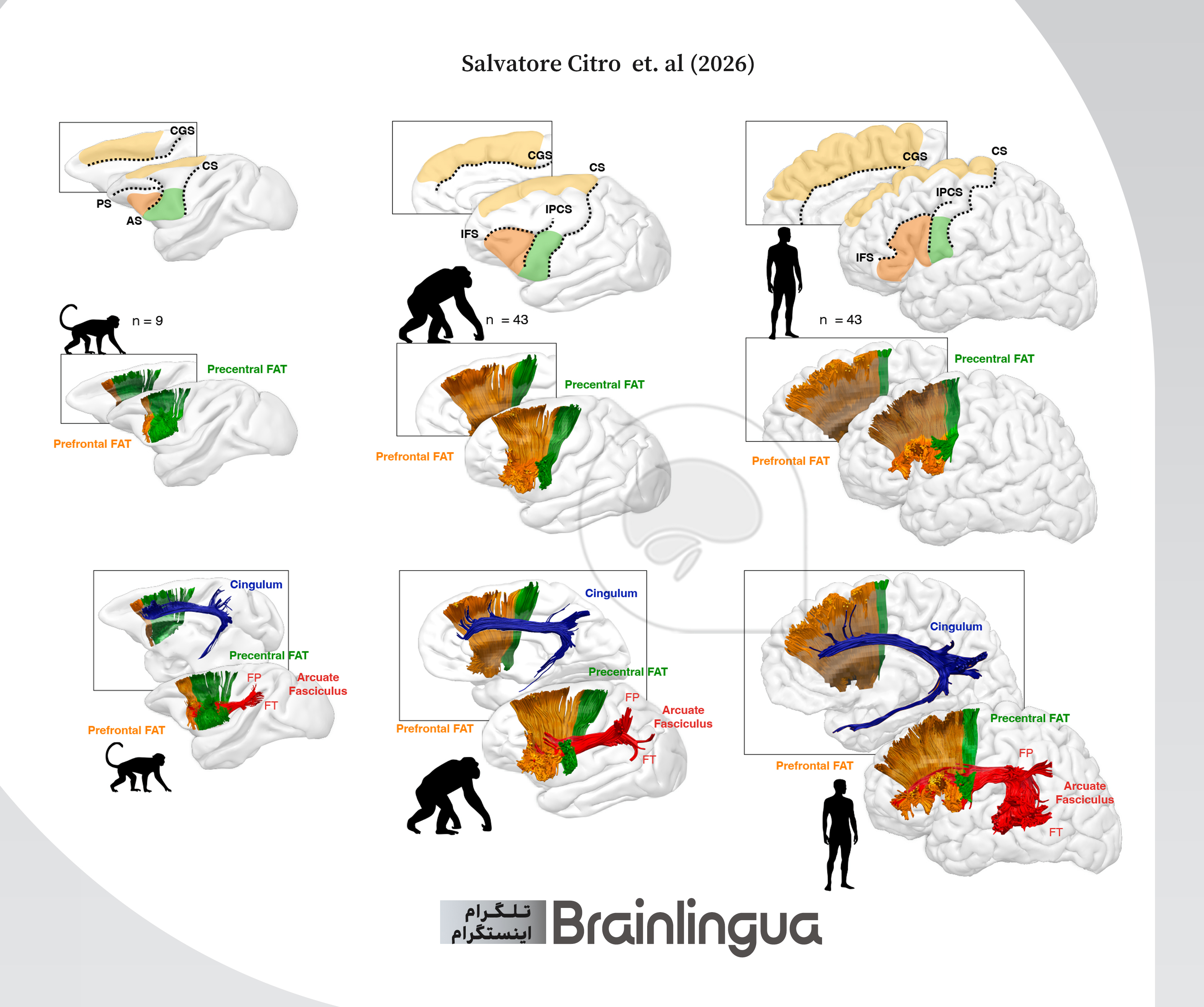
To achieve high resolution in mapping brain networks, diffusion imaging and spherical deconvolution-based tractography were employed, enabling precise tracking in regions with crossing fibers. Neurological data from 9 Old World monkeys, 43 chimpanzees, and 43 healthy humans were extracted and analyzed. Based on anatomical landmarks, the FAT pathway in these species was delineated into two distinct components: posterior (precentral) and anterior (prefrontal). In the clinical phase of the study, data from 98 individuals diagnosed with PPA were investigated. Here, the prefrontal segment of the FAT was further parcellated into three finer subdivisions based on its connectivity with the Inferior Frontal Gyrus (IFG): the pars orbitalis, pars triangularis, and pars opercularis. Subsequently, microstructural diffusion metrics, including Fractional Anisotropy (FA), were calculated. Spontaneous speech capacity was assessed using the Words Per Minute (WPM) metric during the Cinderella storytelling task, while the NAVS-SPPT and NAT assessments were utilized to measure syntactic production capacity and sentence constituent sequencing.
The findings indicate that the proportional total volume of the FAT in the human brain is significantly larger than in monkeys and chimpanzees. However, sub-segmental analyses reveal two distinctly divergent evolutionary trajectories: the precentral FAT in humans and chimpanzees has decreased in size compared to monkeys. In contrast, the prefrontal FAT has undergone substantial expansion; its relative volume constitutes 14% of the entire FAT in monkeys, 67% in chimpanzees, and up to 86% in humans. While the FAT exhibits near-symmetry between the two cerebral hemispheres in monkeys and chimpanzees, this network in humans—particularly within its prefrontal section—demonstrates a highly pronounced left-hemispheric lateralization. This structural asymmetry is considered one of the most unique hallmarks of human brain evolution.
An examination of the input pathways to the FAT reveals that the volume of the cingulum, which relays limbic system information for innate vocalizations, has remained conserved across all three species. Conversely, the arcuate fasciculus, which transmits complex auditory and visuospatial inputs to the ventrolateral frontal cortex, is considerably more massive in humans than in non-human primates. This expansion is exclusively observed in the fronto-temporal segment of the arcuate fasciculus. Furthermore, the analysis of PPA patient data uncovers a clear anatomo-functional gradient within Broca's area and its interconnected pathways. Deficits in speech fluency significantly correlate with reduced white matter integrity in the precentral segment and the posterior portion of the prefrontal cortex (FAT opercularis). Conversely, syntactic processing exhibits a profound dependence on the anterior segments. It was demonstrated that impairments in syntactic production and structural errors in sentence evaluation tasks are directly associated with the degeneration of the FAT orbitalis and FAT triangularis tracts.
This study provides robust evidence in support of the exaptation hypothesis. The FAT network, which in non-human primates served the hierarchical organization of behavior, motor coordination, and the production of instinctive vocalizations, has undergone extensive functional reorganization throughout human evolution. From a cognitive neuroscience perspective, the correlation between syntactic errors and the anterior segments of the FAT (connecting to Brodmann areas BA45 and BA47 in the inferior frontal gyrus) highlights the specialization of these circuits for linear lexico-syntactic processing and semantic-structural integration. The remarkable expansion of the prefrontal segment of this network in humans, coupled with the elaboration of the arcuate fasciculus circuit, has provided the critical neuroanatomical substrate necessary for the precise sequencing of speech sounds and the transition from rudimentary communication to complex syntactic language systems.